Hi, I’m Ciara Doody, a PhD candidate in the St Johnston lab in the Gurdon Institute, University of Cambridge. I’m from a small town in County Galway, in the west of Ireland. I completed my Bachelor’s at the University of Galway, where I studied Biomedical Science, specialising in biochemistry. During my undergraduate degree I became fascinated by cell and developmental biology. I was ready and excited to move to a new place to pursue my PhD, which led me here to Cambridge (which is a lot less rainy than I’m used to!). In my lab we use the fruit fly Drosophila melanogaster to investigate cell polarity.
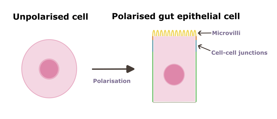
Often images of circles or spheres, apolar shapes, come to mind when we think about cells. In reality, most cells in the body take on very specific shapes in order to carry out tasks. A nerve cell has a long extension used to communicate with other cells. A red blood cell has a dished shape to carry lots of oxygen. The cells that make up the lining of your body are no different. We call these epithelial cells, which act as barriers between the outside and inside of the body. You can these in places such as the skin, respiratory tract and gut. A gut epithelial cell (shown below) has a very distinctive shape, which allows it to fulfil its function. Being a successful gut epithelial cell means knowing where to correctly place the microvilli, cell-cell junctions and correctly orientate absorption and secretion systems. This concept of cells being organised into domains with specific structures and functions is what we call cell polarity. If maintenance of this polarised state is disrupted, cellular activity can become uncoordinated and lead to disease. For example, loss of polarity is associated with metastasis in cancer.
A lot of what we already know about how cells become and remain polarised owes to studies with various tissues in Drosophila, as there is an abundance of experimental tools available within the fly research community. However, what underlies polarity in vertebrate cells (including humans) is less clear. Over the last few years, work in my lab has uncovered that surprisingly, cells in the fruit fly gut seem to polarise in a totally different way than other well-studied fly tissues. In fact, this tissue has many similarities to vertebrate epithelia. This is really exciting because the fruit fly gut may hold the secrets which bridge the gap in our knowledge between vertebrate and invertebrate epithelial tissues. In my PhD project, I am systematically investigating the role of candidate genes in the fly gut, to identify previously undescribed players which take part in the polarisation process. This will hopefully lead to a deeper understanding of one of cell biology’s most fundamental aspects, and in turn, open the door for novel treatments in cases where cell polarity is disrupted.
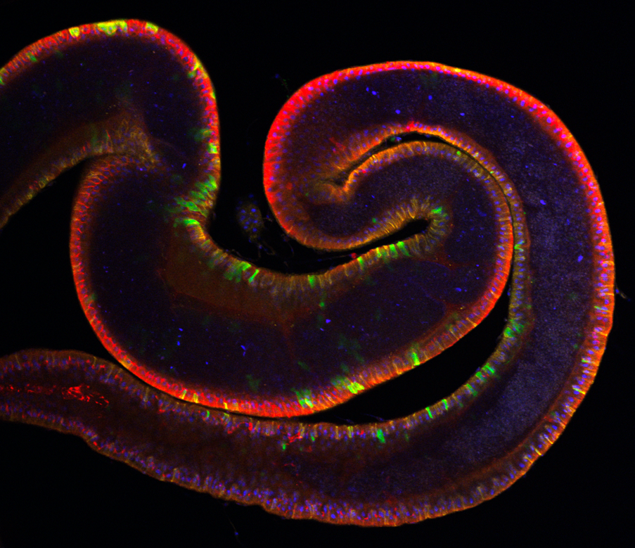
Longitudinal section through a fluorescently labelled fruit fly gut.
In this image, 25% of all cells are labelled with GFP (green fluorescent protein), exemplifying the extent of experimental control we have within the tissue.
